
MEASURED PERFORMANCE
52s
MEDIAN · EDITOR → SIGNATURE
From editor to signature
Historical production snapshot

Fixed snapshot from April 9 to May 9, 2026 · does not measure total TAT or clinical quality. Sample and definitions are available in the methodology.

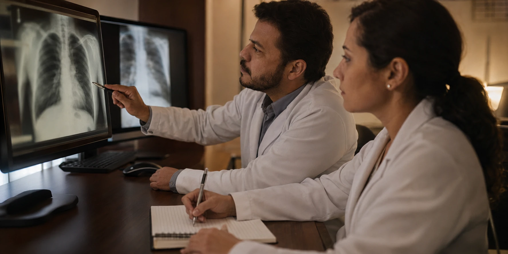
Two radiologists evaluating the same exam
Fidelity in a controlled set

90% score in the v3.10 snapshot of 120 cases. It is not external clinical validation.

Radiologist at a reporting workstation with exams on diagnostic monitorsRadiology managers in a meeting in front of an indicator dashboard
You are
A platform exclusively for healthcare professionals and institutions.

Report far better and far faster.

This is the post-imaging layer of Brazilian radiology: structured reporting that runs from dictation to the signed report and back into the RIS and the PACS of the institution, with no retyping.

A platform exclusively for healthcare professionals and institutions.
Manage radiology with far more intelligence.

Brazilian radiology's post-imaging layer: throughput, TAT and critical findings with an SLA, in your RIS and your PACS.

52s
median
Measured performance
From editor to signature
Result measured in productionSee results
Try it in your real routine14 days free or up to 30 reports
Your exams, your templateon your own shift, not a demo environment
No credit cardno automatic charge
After the trial, if it makes sensePro: R$ 219/month, up to 1,000 reports
FILM · 77 SECONDS

The post-image layer in 77 seconds

Speak and the template fills in. Describe and the classification is there. Edit by voice, report multiple exams in one recording and leave a trail in the workflow, with CFM Resolution 2.454/2026 along the way.

  1. The image, you have already solved. The report is still blank.
  2. Speak. Your template fills in.
  3. Not transcription. Writing.
  4. You describe. The classification is there.
  5. Critical finding in the text. Alert on the spot.
  6. Edit by voice. Multiple exams, one recording.
  7. Made by those who report.
  8. Enters the workflow. Leaves a trail.
  9. CFM 2.454 compliance.
  10. Not a promise. A measurement.
  11. You review.
  12. For everything that comes after the image.
ASSISTIVE AI THE GOVERNED FLOW

You gain speed.
The report gains a standard.

You need to cut repetitive tasks and spend more time on diagnosis, without giving up your institution's standard. In production, that is close to 10 radiologist hours recovered per 100 reports — and it is the same platform that gives the time back and shows where it went.

Context ready before the first click
SOURCES
Worklist · indication · priors
ORIGIN
RIS / PACS, with no manual upload
Live structuring, review visible throughout +
STRUCTURE
Technique · findings · impression
CONTROL
Zero-Click: review visible throughout
Critical findings and audit trail +
CRITICAL
SLA + escalation (CRIT)
AUDIT
Auditable log per exam
AUDIO · CONFIGURABLE PROCESSING DATA · DISTINCT LIFECYCLES
Data flow documented for each deployment

Audio, transcription, draft, final report, metadata, logs, and backups are distinct categories. Transmission, persistence, and retention depend on the feature and contracted configuration.

REGION AND SUBPROCESSORS · DOCUMENTED
Radiology reporting workstation with a dictation microphone and images on monitors
CT
Dr. Marina Duarte
Chest CT · 09:30
EXAM CONTEXT

Non-contrast chest CT to investigate a chronic cough of 3 months. Former smoker, 20 pack-years. May radiograph with a right upper lobe opacity for correlation. 2024 prior in the PACS; indication and history imported from the RIS.

3 sources · PACS/RIS
Dictated findings
LIVE
Ground-glass opacity
Right upper lobe, 8 mm
Mediastinal lymph nodes
Calcified
2024 prior
Stable in size
No effusion or pneumothorax
Osseous structures preserved
REC 12:04 ❚❚ PAUSE
  1. BEFORE THE EXAM
    The radiologist interprets

    You keep your natural flow: look at the image, decide and dictate — no commands, no memorized phrases.

  2. DURING DICTATION
    The AI keeps up

    It structures, organizes and accelerates: the report assembles in your house style in real time while your eyes stay on the image.

  3. AFTER THE SIGNATURE
    The institution follows along

    You review and sign — and indicators, quality and performance stay visible, with an auditable trail. More productive physicians, smarter institutions, better-served patients.

REAL CASES

The dictation as you give it.
The report that comes out.

Running speech, with pauses, mid-sentence corrections and on-call slang. On the right, the text that reaches you to review, edit and sign.

DICTATED SPEECH

“Look, there are findings suggestive of pulmonary embolism, yes. It is saddle there, so central PE, right? And besides that I am seeing an aerated consolidation here in the posterior basal segment of the right lower lobe, about 6.3 centimeters. Since it is in the setting of PE, I think it could be an infarction.”

“And wow, this interventricular septum is bulging too, that could even point to right heart strain. At the thoracoabdominal transition just signs of a prior cholecystectomy and bilateral calyceal microcalculi, about two on each side, up to 0.3, non-obstructive. That is all, all right?”

GENERATED REPORT · CHEST CT ANGIOGRAPHY

FINDINGS

Pulmonary trunk and main pulmonary arteries: vascular filling defect extending through the bifurcation of the pulmonary trunk and the main pulmonary arteries (saddle pulmonary embolism).

Cardiac chambers: bulging of the interventricular septum, indicating signs of right heart strain.

Lung parenchyma: consolidation with air bronchograms in the posterior basal segment of the right lower lobe, extending 6.3 cm, suggestive of pulmonary infarction.

Thoracoabdominal transition: prior cholecystectomy. At least two calyceal microcalculi on the right and two on the left, measuring up to 0.3 cm, non-obstructive.

IMPRESSION

Signs of acute central (saddle) pulmonary embolism, with right heart strain and consolidation in the right lower lobe consistent with pulmonary infarction.

CRIT Acute central pulmonary embolism flagged as a critical finding, for recorded review and communication.

DICTATED SPEECH

“This is a thyroid ultrasound. The gland is in place, normal dimensions, texture a little heterogeneous. And most importantly: a solid hypoechoic nodule in the middle third of the right lobe, measuring 1.2 cm. Wider than tall, regular margins, no microcalcifications. No lymphadenopathy.”

ADDITION DICTATED LATER

“I would like you to add, man, something I forgot to say: there is a really hypoechoic lesion below the right lobe, it looks like the matching parathyroid. It measures 1.2 cm and on Doppler it has a somewhat odd vascularity, with a pedicle. Could it be a lymph node? It could. But I do not know exactly what it is.”

GENERATED REPORT · THYROID ULTRASOUND

FINDINGS

A solid, hypoechoic nodule is identified in the middle third of the right lobe, with regular margins, wider than tall and without microcalcifications, measuring 1.2 cm (ACR TI-RADS 4).

Solid, markedly hypoechoic nodular formation underlying the lower pole of the right lobe (topography of the right inferior parathyroid), measuring 1.2 cm, with a probable polar vascular pedicle on Doppler. The appearance suggests an enlarged parathyroid; an exophytic thyroid nodule or atypical lymphadenopathy should be considered in the differential diagnosis.

IMPRESSION

Solid hypoechoic nodule in the right thyroid lobe (ACR TI-RADS 4) and a vascularized nodular formation underlying the lower pole, suggestive of an enlarged parathyroid, to be correlated with serum calcium and PTH.

GUIDE The ACR TI-RADS criterion arrives alongside the finding, with the version cited. Category and management are medical decisions.

DICTATED SPEECH

“This is a multiparametric prostate MRI. Prostate with increased volume, 45 grams. In the right posterolateral peripheral zone, middle third, there is a spiculated nodule with T2 hypointensity, marked diffusion restriction, a drop on the ADC map, measuring 1.6 centimeters.”

“There is early contrast enhancement, important. And it is touching the capsule, with slight bulging, but no seminal vesicle invasion and no lymphadenopathy in the pelvis. The transition zone is just the usual benign prostatic hyperplasia.”

GENERATED REPORT · MULTIPARAMETRIC PROSTATE MRI

FINDINGS

Peripheral zone: nodular lesion with spiculated contours in the middle third of the right posterolateral peripheral zone, with T2 hypointensity, marked diffusion restriction with corresponding drop on the ADC map and early contrast enhancement, measuring 1.6 cm along its greatest axis.

Prostatic capsule: contact and slight bulging adjacent to the described nodule, without definitive evidence of gross extraprostatic extension. Seminal vesicles with no signs of invasion. No pelvic lymphadenopathy.

IMPRESSION

Lesion with a high probability of clinically significant prostate cancer (PI-RADS 5), causing slight focal capsular bulging. Signs of benign prostatic hyperplasia in the transition zone.

GUIDE The PI-RADS v2.1 category is proposed with the criterion alongside it and the reference versioned in RadCommons. You are the one who validates it.

Examples of dictation and drafts generated on the platform, with identifying data removed. The AI is assistive: the radiologist provides the findings, reviews, edits and signs. See the interactive demonstration.

Science and compliance

Artificial intelligence is only the beginning.

The goal is to organize the post-imaging layer with fewer operational steps and explicit controls. The physician provides the findings, reviews, edits, validates, and signs; the data flow is documented for each deployment.

Resolution CFM 2.454/2026
Personal data LGPD ANPD
01
90% Agreement with the physician's report
Hundreds of real reportsLaiBench v3.10
LaiBench

90% match against the signed report

We ran hundreds of real reports and compared them with what Laudos.AI would write. Nine out of ten came out identical to what the physician signed — and LaiBench measures that on every version.

02
Test dateJuly 2026
Version testedLaiBench v3.10
Number of reportsHundreds of real cases
Who signsThe physician, every time
Methodology

Open number, math on the table

When the test ran, which version went in, how many reports were measured. You see the whole methodology — few show it.

03
v4Pulmonary noduleSource cited
v2Carotid stenosisSource cited
v1Breast densityUnder review
RadCommons

No score leaves without a source

Every classification comes with the reference that supports it and the version of the criterion. You sign knowing exactly where each number came from.

04
What changed in the review
3.2 cm nodule in the right upper lobe
3.8 cm nodule in the right upper lobe
Reviewed and signed by the physician2nd version of the report 14:38
Audit

You in control, every time

Laudos.AI writes the draft, you adjust it and sign. Every change is recorded with author and time — audit solved before it turns into a problem.

05
CFM 2.454/2026 LGPD · ANPD
Rule announced
1st week
100% compliant
CFM 2.454/2026 · LGPD

Compliant from the first week

CFM 2.454/2026 was announced and Laudos.AI was already 100% compliant in the first week. LGPD, the same: patient data handled within the law, with no compliance project halfway through.

COMPARISON

The usual workflow and the post-image layer, criterion by criterion

By what changes in the routine, not by the feature list. The Laudos.AI numbers are the ones measured in production and described in the methodology.

CriterionManual workflowLaudos.AI
Time from editor open to signatureClose to 10 minutes per report, in the literatureMedian of 52 seconds; 54,6% of reports in under 1 minute, measured in production
Report structureThe house template lives in a shared folder and ages on its ownTechnique, Analysis and Impression assemble in the house template while you dictate
ClassificationThe classification memorized or open in another window, in the middle of the reportGUIDE proposes the category with the current criterion visible, for medical review
Critical findingA phone call with no record, no SLA and no proof that anyone answeredA protocol with level, SLA, escalation and closed loop
AuditThe trail is rebuilt record by record when a request is madeOne log per exam reconstructs who saw, what changed and when
Return to the RIS and the PACSCopy and paste between the editor and the system, every dayThe signed report returns to the house RIS and PACS, in the format they already accept

The manual workflow time comes from the peer-reviewed literature cited in the references on this page. See the comparisons by criterion →

FIG. 01 · LAIBENCH
Fidelity measured in a controlled internal set.

The v3.10 snapshot records a 90% score across 120 cases. It is a fidelity benchmark, not external clinical validation.

See LaiBench
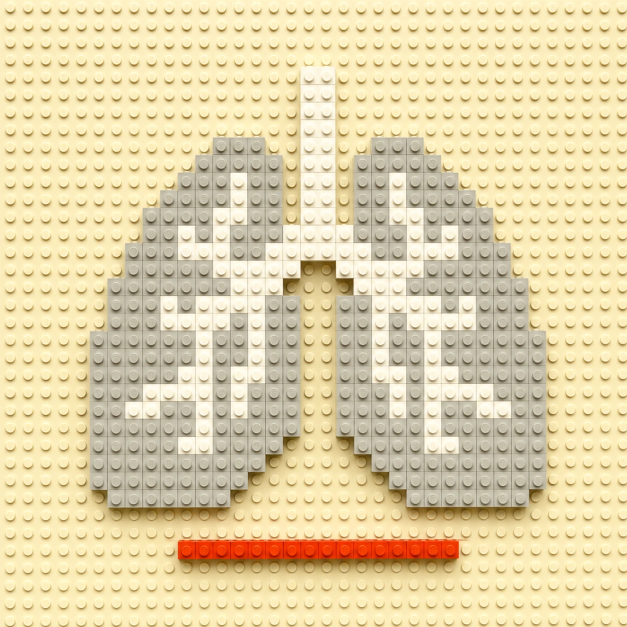
Fig. 01 · LaiBench v3.10 snapshot
CT scanner with its cover open in an exam room
laudos.ai Manifesto
Everything that comes after the image.

Artificial intelligence does not replace specialists — it lets them do more. The future of radiology is not human versus machine: it is physicians and technology together for better medicine.

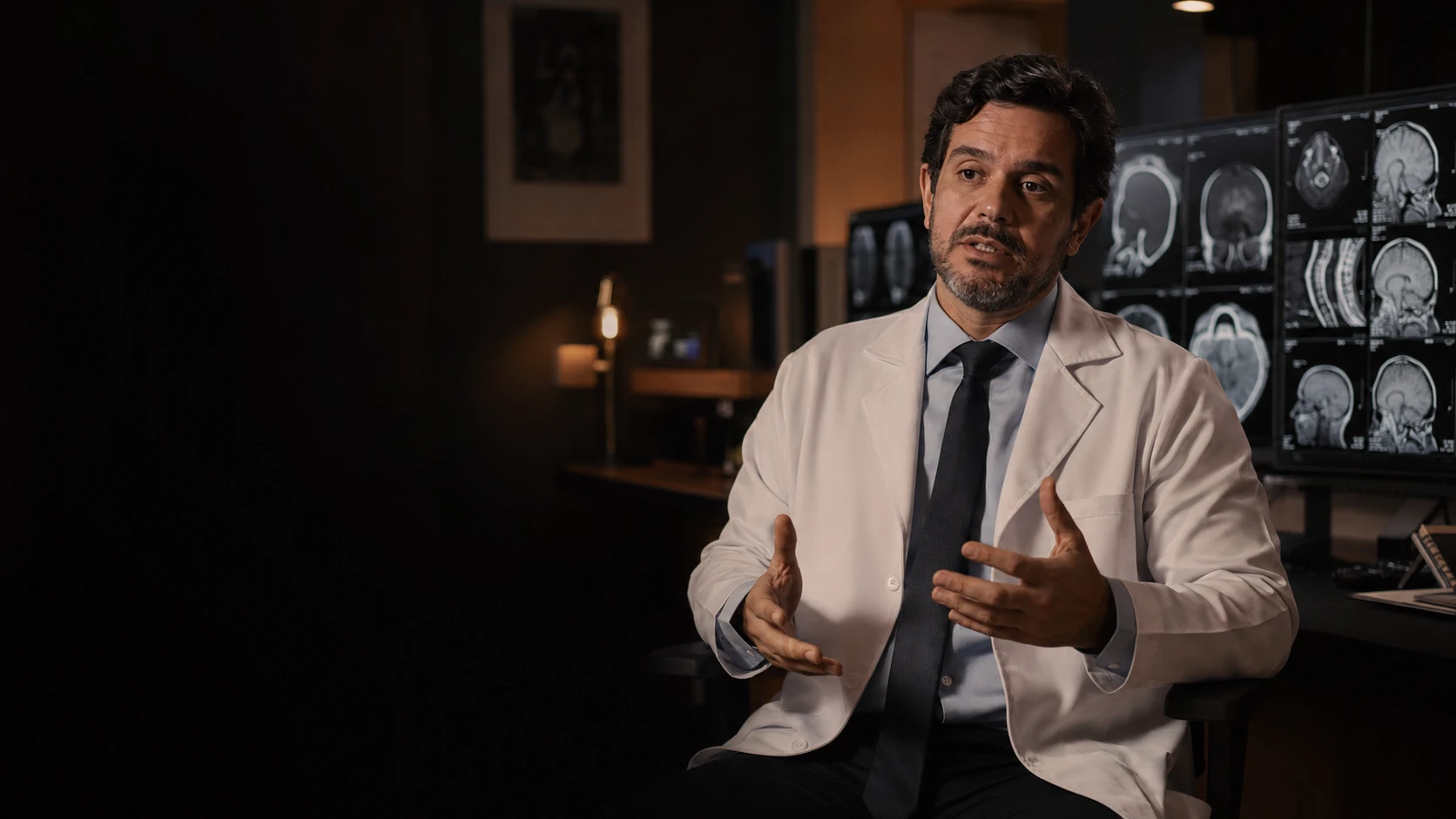
Radiologist being interviewed next to imaging monitors
Far beyond faster reports: the time goes back to diagnosis.

Close to 10 radiologist hours recovered per 100 reports, measured in production. The report comes out in your institution's format, the classification arrives with the criterion beside it and the critical finding travels with a record — without taking your eyes off the image.

RADIOLOGY · LIVE
REAL-TIME TAT SLA MONITORED
DEPLOYMENT

From first contact to the real flow

  1. 01
    A demo in your context

    Your exam types, your institution’s template, your flow. No slides, straight into the product.

  2. 02
    A pilot with the team

    Radiologists using it in a real routine, with the institution’s templates, before any decision is made.

  3. 03
    Assisted integration

    PACS, RIS and worklist connected. The signed report goes back into your system.

Security documentation ready for IT and DPO review: architecture, encryption, subprocessors and data flow.

WHO USES IT

Radiologists who leave the shift
with less friction.

  • Laudos.AI helps me move faster on the cases I know well and describe less familiar findings with more structure. I cannot imagine working without it.
    Dra. Stephanie A.MSK radiologist · CRM-SP 192818
  • I use Laudos.AI for my Dopplers. The precision in structuring the findings is what stands out most.
    Dr. Bernardo HimeVascular surgeon · CRM-SP 192561
  • As a first-year resident, I do not know what my routine would be without it. I dictate a finding and get back a structured description that makes sense.
    Dr. Petrus ParaísoRadiology resident
  • The reporting assistant has helped me a lot, not only in speeding up reports but also in feeling more confident that I am conveying the information clearly. I particularly like the diagnostic impressions it generates.
    Dra. TabitaRadiologist · Fortaleza, Brazil
  • Your platform created a real disruption. We are not talking about a simple transcription tool, but about an outstanding way to organise your thinking and structure the report.
    Dr. Fábio BortoliniRadiologist
  • I can report every exam within the shift itself. I no longer take any work home. And it prevents rework too.
    Dr. Carlos IlhaRadiologist · Paraná, Brazil
DIRECT QUESTIONS

Before you
book

Who is the author of the report?

The radiologist, every time. The AI is assistive: it structures and proposes. Review, editing and signature are the physician’s.

Is my data used to train models? +

Identifiable healthcare data is not used to train general-purpose models. Processing, persistence, logs, retention, and backups are addressed separately in the documentation for the contracted feature.

Where does the data live? +

The region varies by feature and subprocessor. The applicable flow, including any international transfer, is presented to the institution for assessment.

Does it integrate with my PACS and RIS? +

It depends on the system, version, and connector. API, HL7/DICOM, and Agent are distinct paths validated during mapping; we do not promise universal compatibility.

And our own vocabulary? +

Institutional templates per modality, with governance and versioning: your institution’s model is applied to every report.

Do I need to change my workflow? +

The pilot maps templates, permissions, connectors, and contingency. The degree of workflow change depends on the current environment and the modules adopted.

How are critical findings handled? +

CRIT is in a controlled pilot.

  • Detection depends on the findings provided by the physician.
  • Confirmation, channels, recipients, timeframes, and closure are defined and tested for each deployment.
What is the regulatory status? +

A medium-risk assistive application under CFM Resolution 2,454/2026. It is not SaMD; it does not autonomously interpret images, diagnose, sign, or release reports. The physician provides the findings, reviews, edits, validates, and signs in the official system. See the control matrix.

How does deployment work? +

Within days your team is up and reporting.

  1. Workflow mapping (PACS/RIS, modalities, and templates).
  2. A demo with synthetic cases.
  3. A pilot with baseline metrics.
  4. Monitored production with support and auditing.
What if the AI is wrong or unavailable? +

Incorrect suggestions are to be edited or discarded by the physician. During unavailability, the institution's contingency plan applies; we do not presume every workflow continues without impact. The public status shows only dated checks.

How do the trial and the subscription work? +

You try it for 14 days or up to 30 reports, no card and no automatic charge. If it makes sense to continue, Pro costs R$ 219/month for up to 1,000 reports — close to R$ 0.22 per report. See the details at /precos.

WHO IT SERVES

Different roles,
explicit controls.

Use scenarios, not testimonials. The radiologist remains responsible for interpretation and the report; the institution governs deployment, access, integration, and contingency.

RADIOLOGIST
From dictation to a structured draft

Provides findings and retains full control over review, editing, validation, and signing.

CLINICAL MANAGEMENT
Templates and governance

Defines standards, permissions, indicators, and contingency criteria for the local context.

IT AND DPO
Verifiable deployment

They assess connectors, data flows, subprocessors, access, retention, and evidence before production.

REFERENCES
  1. European Society of Radiology (ESR). ESR paper on structured reporting in radiology. Insights Imaging. 2018;9(1):1-7. doi:10.1007/s13244-017-0588-8
  2. Ringler MD, Goss BC, Bartholmai BJ. Syntactic and semantic errors in radiology reports associated with speech recognition software. Health Informatics J. 2017;23(1):3-13. doi:10.1177/1460458215613614
  3. Hawkins CM, Hall S, Hardin J, Salisbury S, Towbin AJ. Prepopulated radiology report templates: a prospective analysis of error rate and turnaround time. J Digit Imaging. 2012;25(4):504-511. doi:10.1007/s10278-012-9455-9
  4. Larson DB, Towbin AJ, Pryor RM, Donnelly LF. Improving consistency in radiology reporting through the use of department-wide standardized structured reporting. Radiology. 2013;267(1):240-250. doi:10.1148/radiol.12121502
OPERATIONAL LIBRARY · CFM 2.454/2026

The question is proving the AI was used correctly.

CFM Resolution 2.454/2026 comes into force on 26 August 2026 and demands evidence produced inside the workflow, not internal policy. It is 16 pages, six playbooks and 36 implementation steps, sourced only from official standards: CFM, LGPD, ANPD, ACR, IHE, HL7, DICOM and NIST.

01
Auditable record

What has to be recorded per exam, and in what format, to hold up in an audit.

02
Demonstrable clinical supervision

How to turn medical review into evidence, instead of a statement of intent.

03
Preserved medical authorship

Who signs, what changed after the suggestion and how that is recorded.

NEXT STEP

More intelligence for every report.

Start reporting with REPORT, GUIDE and CRIT in your real routine.

Individual plan: 14 days or 30 reports, no card.

More control across the entire operation.

Bring REPORT, GUIDE, CRIT, structured data and an audit trail to your institution.

Demo with synthetic data and pilot design.
Report faster.
REPORT, GUIDE and CRIT in your routine
Start the evaluation

FAQ

Questions and answers

Individual plan: 14 days or 30 reports, no card.

What it is
The post-imaging layer of Brazilian radiology: from dictation to the structured report, back into the RIS and PACS.
Who it is for
Radiologists who report and institutions: clinic, hospital and teleradiology.
What it measures
Median of 52 seconds from editor open to signature; 54.6% of reports under one minute; close to 10 hours returned per 100 reports.
What it does not do
It does not interpret images, does not diagnose, and does not sign or release reports autonomously.
In summary
Structured reporting from dictation to signature, back into the RIS and the PACS, with an audit trail per exam; the AI does not sign in the physician's place.

Figures published on this page

Median from editor open to signature
52 s
April 9 to May 9, 2026 · 5,200 finalized reports
Metrics methodology
Reports under one minute
54.6%
Same window
Metrics methodology
Radiologist hours returned
Close to 10 per 100 reports
Same window
Metrics methodology
LaiBench v3.10 · fidelity score
90% on 120 cases
Controlled internal set · June 23, 2026
LaiBench
Resolution 2.454/2026
In force from August 26, 2026
Published on February 27, 2026
Operational Library for CFM 2.454
Pro plan
R$ 219/month for up to 1,000 reports
14-day or 30-report trial, no card
Pricing

Product

What is Laudos.AI?

Laudos.AI is the post-imaging layer of Brazilian radiology: it turns dictation into a structured report and returns it to the RIS and the PACS, with a median of 52 seconds from editor to signature, measured in production.

  • For radiologists who report and for clinics, hospitals and teleradiology.
  • The AI is assistive: it structures and proposes; review, editing and signature are the physician's.

Product

What does structured report mean at Laudos.AI?

Structured reporting, at Laudos.AI, refers to the report text organized into fixed sections, with findings, measurements and classifications in the right fields, generated from free dictation. This means that the physician dictates the way they reason, the AI organizes the text, and interpretation stays with the physician. Product

What are the platform's modules?

REPORT in production; GUIDE and LaudAI in beta; CRIT in a controlled pilot; Agent and the institutional dashboard in beta.

  • REPORT: the editor that structures the report from dictation.
  • GUIDE: BI-RADS, LI-RADS, PI-RADS and other classifications at the point of the report.
  • CRIT: critical findings with identification, physician confirmation and recorded communication.
  • LaudAI: the report in the institution's standard, back into the RIS and PACS.

Modules

Does the platform interpret images?

No: interpretation belongs to the physician. The platform structures the text from the dictated findings and proposes classifications.

  • The physician provides the findings; the AI structures the text and proposes classifications.

Compliance

Does the AI sign the report?

No: the physician always signs. The AI structures the text and proposes; signature and release stay in the official system.

  • The platform does not interpret images or make autonomous diagnoses.
  • The physician provides the findings, reviews, edits, validates, and signs in the official system.

Compliance

What is a governed critical finding?

A governed critical finding refers to a result that has to be communicated within a deadline, with the physician in control: CRIT asks for the radiologist's confirmation, communicates with an SLA per level and records each step in the exam's audit trail. As stated on the product page, CRIT is in a controlled pilot. Product

How does the flow from dictation to the RIS work?

How it works, in five steps: the worklist, the indication and prior exams arrive from the RIS and the PACS with no manual upload; the radiologist dictates; the AI structures the text and proposes classifications; the physician reviews, edits and signs in the official system; the report returns to the RIS and the PACS with no retyping. According to the integrations page, that return is validated per deployment. Integrations

Does it integrate with my PACS and my RIS?

Yes: through API, HL7, DICOM and the Agent, with the report's return validated in your deployment.

  • Compatibility depends on the system, version, connector, and contracted scope.
  • Return to the RIS or PACS is validated in each deployment, so there is no universal integration promise.

Integrations

What is RadCommons?

RadCommons is defined as Laudos.AI's repository of classification systems: 121 versioned systems, with cited sources and served over an API. In the editor, the category is shown with the criterion alongside it and the versioned reference; the radiologist validates it. RadCommons

Pricing and deployment

What does it cost?

R$ 219 per month on the individual plan, with up to 1,000 reports a month, close to R$ 0.22 per report; institutions receive a proposal sized by exam volume and active modules.

  • 14-day or 30-report trial, no card.
  • No hidden per-user fee.

Pricing

Is there a trial before subscribing?

Yes: 14 days or 30 reports, whichever comes first, with no card and no automatic charge.

  • Up to 5 of your own templates during the trial.

Start the trial

How long does deployment take?

Up to two weeks, in three phases, without replacing the PACS or the RIS.

  • The pilot maps templates, permissions, connectors and contingency.

Deployment

Does it work for clinics, hospitals, and teleradiology?

Yes, and also for the radiologist who reports alone: an individual plan with public pricing and an institutional proposal sized by exam volume and active modules.

  • Radiologist reporting solo: individual plan with a public price and a new-user trial with no card required.
  • Clinic, hospital, and teleradiology: a proposal by exam volume and active modules, with integration validated for each deployment.

Solutions by context

Bottom line: what does Laudos.AI deliver?

In summary: structured reporting from dictation to signature, back into the RIS and the PACS, with governed critical findings, operational data and an audit trail per exam: measured in production, with the physician signing every time. Product

Figures

Where does the 52-second figure come from?

52 seconds is the median, measured in production, from opening the editor for an exam to signing the report.

  • Fixed historical snapshot: April 9 to May 9, 2026.
  • Sample: 5,200 completed reports.
  • It does not measure total service TAT, clinical quality, causality, or future performance.

Metrics methodology

What is the difference between median and mean reporting time?

The 52-second median is the typical report; the mean, between 3 and 5 minutes, includes the complex cases. Laudos.AI publishes both.

  • 54.6% of reports come in under one minute in the same measurement.
  • We publish median and mean side by side so the tail is not hidden.

Metrics methodology

Who measures the figures published on this page?

Laudos.AI itself, in production, with a public methodology. According to the methodology page, the window is fixed, April 9 to May 9, 2026, with 5,200 finalized reports; according to the same page, the figure measures neither the total TAT of the service nor clinical quality. Metrics methodology

What is TAT, and how does it differ from the 52 seconds?

TAT, also known as turnaround time, refers to the total time from the exam being performed to the report being released, and it is shown on the institutional dashboard; the 52 seconds measure only the stretch from editor open to signature, the part the radiologist performs inside the platform, because that is where the platform acts. This means the two figures are neither added together nor compared. Metrics methodology

How do you prove that quality holds up?

With a published benchmark: LaiBench v3.10, a fidelity score of 90% on 120 cases from a controlled internal set, on June 23, 2026.

  • It is a fidelity benchmark, not external clinical validation.
  • The radiologist remains responsible for interpretation and the report.

LaiBench

What is LaiBench?

LaiBench can be described as Laudos.AI's fidelity benchmark, published with the math on the table: it compares the text structured by the AI against the signed report in a controlled internal set. According to the LaiBench page, v3.10, from June 23, 2026, scored 90% on 120 cases. It is not external clinical validation, because the set is internal and controlled. LaiBench

How do I compare Laudos.AI with other reporting platforms?

By three measures that Laudos.AI publishes and measures in production: time from editor to signature, return to the RIS and the PACS with no retyping, and an audit trail per exam.

  • Time from open editor to signature, measured in production.
  • Report return to the RIS and PACS without retyping.
  • Per-exam audit trail and compliance with CFM Resolution 2,454/2026.

Compare reporting platforms

Compliance and data

What changes with CFM Resolution 2.454/2026?

Laudos.AI is classified as an assistive application of medium risk, not as Software as a Medical Device; human supervision, medical authorship and traceability are already part of the product.

  • Its controls support human oversight, physician authorship, and traceability.
  • Final compliance also depends on the deployment context and the responsibilities of the institution and physician.

Operational Library for CFM 2.454

When does CFM Resolution 2.454/2026 take effect?

Published on 27 February 2026, it takes effect on 26 August 2026.

  • The application is assistive, classified as medium risk, and is not Software as a Medical Device.

Operational Library for CFM 2.454

What does CFM Resolution 2.454/2026 require of a reporting AI?

Meaningful human supervision, medical authorship and traceability, according to the CFM 2.454 Operational Library kept on this site. Laudos.AI is classified as an assistive application of medium risk, not as Software as a Medical Device, which is why the AI neither signs nor releases reports and each exam keeps an audit trail. Operational Library for CFM 2.454

Where is exam data stored?

Each type of data follows a flow documented per deployment, defined by the feature and the contracted configuration.

  • Audio, intermediate transcription, draft, final report, logs, and backups have distinct flows and retention periods.
  • Some subprocessors may process data outside Brazil.

Security

Does patient data train models?

No: identifiable care data does not train general models.

  • The processing needed to deliver the contracted function is a separate purpose and is recorded in the deployment documentation.

Security

Is there an audit trail per exam?

Yes: each exam produces audit records proportional to the contracted function, validated before production.

  • Before production, deployment validates the trail's events, content, retention and availability.

Compliance

Try it in your real routine

14 days free or up to 30 reportsNo credit card

Content updated on .